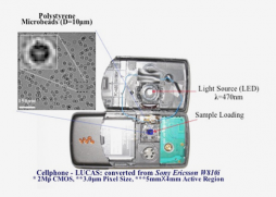
Wired has finally picked up the story that has been circulating for a while -- the phenomenal medical diagnostic hack using a mobile and beginning to turn it into a lab for developing countries.
Aydogan Ozcan, assistant professor of electrical engineering at the UCLA School of Engineering and Applied Science and a member of the California NanoSystems Institute (CNSI), and his team of graduate and udergraduate students developed a medical diagnostic application from a mobile phone, in effect bringing the hospital to the patient.
In developing countries, the distance between people in need of health care and the facilities capable of providing it is a huge obstacle. As E-health wrote in September, Ozcan developed a "novel lens-free, high-throughput imaging technique for potential use in such medical diagnostics, which promise to improve global disease monitoring'.
Or, as Wired put it a bit more catchily:
A new MacGyver-esque cellphone hack could bring cheap, on-the-spot disease detection to even the most remote villages on the planet. Using only an LED, plastic light filter and some wires, scientists at UCLA have modded a cellphone into a portable blood tester capable of detecting HIV, malaria and other illnesses. Blood tests today require either refrigerator-sized machines that cost hundreds of thousands of dollars or a trained technician who manually identifies and counts cells under a microscope. These systems are slow, expensive and require dedicated labs to function. And soon they could be a thing of the past.
UCLA researcher Dr. Aydogan Ozcan images thousands of blood cells instantly by placing them on an off-the-shelf camera sensor and lighting them with a filtered-light source (coherent light, for you science buffs). The filtered light exposes distinctive qualities of the cells, which are then interpreted by Ozcan's custom software. By analyzing the cell types present in a much larger sample, a more accurate diagnosis can be made in a matter of minutes. No more sending blood away to a lab and waiting days or weeks for the results.
The research - online here -- describes improvements to a technique known as LUCAS, or Lensless Ultra-wide-field Cell monitoring Array platform based on Shadow imaging. E-health writes:
First published in the Royal Society of Chemistry's journal Lab Chip in 2007, the LUCAS technique, developed by UCLA researchers, demonstrated a lens-free method for quickly and accurately counting targeted cell types in a homogenous cell solution. Removing the lens from the imaging process allows LUCAS to be scaled down to the point that it can eventually be integrated into a regular wireless cell phone. Samples could be loaded into a specially equipped phone using a disposable microfluidic chip.
The UCLA researchers have now improved the LUCAS technique to the point that it can classify a significantly larger sample volume than previously shown up to 5 milliliters, from an earlier volume of less than 0.1 ml representing a major step toward portable medical diagnostic applications.
Ozcan envisions people one day being able to draw a blood sample into a chip the size of a quarter, which could then be inserted into a LUCAS-equipped cell phone that would quickly identify and count the cells within the sample. The read-out could be sent wirelessly to a hospital for further analysis. "This on-chip imaging platform may have a significant impact, especially for medical diagnostic applications related to global health problems such as HIV or malaria monitoring," Ozcan said.
LUCAS functions as an imaging scheme in which the shadow of each cell in an entire sample volume is detected in less than a second. The acquired shadow image is then digitally processed using a custom-developed "decision algorithm" to enable both the identification of the cell/bacteria location in 3-D and the classification of each microparticle type within the sample volume.
Various cell types such as red blood cells, fibroblasts and hepatocytes or other microparticles, such as bacteria, all exhibit uniquely different shadow patterns and therefore can be rapidly identified using the decision algorithm....
"This is the first demonstration of automated, lens-free counting and characterization of a mixed, or heterogeneous, cell solution on a chip and holds significant promise for telemedicine applications," Ozcan said."This is a significant advance in the quest to bring advanced medical care to all reaches of the planet," said Leonard H. Rome, interim director of the CNSI and senior associate dean for research at the David Geffen School of Medicine at UCLA. "The implications for medical diagnostic applications are in keeping with CNSI initiatives for new advances toward improving global health."
The full study is here, and the fabulous slideshow of the mobile hack is here.


Post new comment